“Wow! There it is, I see it!”
By Dr. Andrea Honigmann, DVM
May 7th, 2024
Walking into a cavernous room with a very visible pulley system overhead, I headed to the back of the room where there were tables with microscopes. It was the fall of 2001 and my first year of veterinary school at Iowa State University, and this was histology class. Histology, the microscopic anatomy of biological tissues, meant there was a lot of microscope time when not in lecture. According to the designers of the curriculum, this paired well with the gross anatomy class that we were taking concurrently, and so as a first year student, this laboratory was where I spent most of my days and many late nights.
I distinctly remember using the dual-headed microscopes (where the professor and student can look at the same slide simultaneously) during lab and nodding my head, giving the instructor a positive indication that I was seeing precisely what she wanted me to see. In reality, I felt like Jennifer Anniston’s character “Rachel” in the iconic TV show Friends, where she’s looking at the ultrasound of her baby for the first time. She says to the doctor, “Wow! There it is, I see it!” The doctor leaves the room, and Ross says, “Pretty amazing, huh?” And then Rachel is tearfully honest, and says emphatically, “I don’t see it!!”
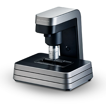
Of course, over the years in veterinary school and beyond, I became proficient at identifying cells of concern on a microscope slide, but it was never really something I enjoyed. What I do enjoy, though, is technology in veterinary practice and how it can make my life easier while allowing me to provide a better service to my clients. While the microscope is still very much a part of veterinary practice, it looks a lot different now than that dual-headed microscope that I learned on as a student in the early 2000s. The microscopes in my practice are digitally connected and about the size of toaster turned upright on its short end. With those small powerhouses I can perform a needle aspirate on a lump or bump on a pet and connect to board-certified pathologists in a matter of minutes. This allows for a diagnostic answer in about 2 hours.
Prior to Zoetis’ IMAGYST and these microscopes arriving in our practice, the same task would be done either in the clinic by myself or sent to a reference laboratory through a courier service. The time at the reference laboratory could be a week to ten days before I could get an answer for an anxious client (when dealing with the potential for cancer in their beloved pets). By having the accessibility through technology like this we can get a diagnosis and an action plan together in a fraction of that time. This leads to treatment plans being developed with my clients sooner and pets getting the care they need much faster.
Not only do these digitally connected microscopes allow me to get cytology answers faster, but with the use of artificial intelligence (AI), I can diagnose parasitic infections, urinary tract infections, ear infections and even review the red and white blood cells of patients, in just minutes. Having these devices in my practice has truly revolutionized the level of care that I can provide to patients. Before this it was only the occasional case where I would walk a client into the treatment area to try to show them what I was seeing on the microscope (and undoubtedly, many of my clients were probably “Rachels,” not really seeing what I saw).
Now, in 2024, I can print out a color image (see above) of exactly what I’m seeing or show the client directly on my laptop in the exam room. Technology is ever-evolving in and out of the veterinary space. Having Zoetis IMAGYST and the AI technology that powers it as part of my toolkit, allows me to practice in a way that levels up the care that I can provide to pets and the humans that love them. The result is that now everyone can see what I see, including Rachel!
Andrea Honigmann, DVM
Hannastown Veterinary Center


